Cardiac Imaging
Cookeville Regional Medical Center
is the only facility in the
Upper Cumberland that offers
cardiac imaging.
From cardiac CT, cardiac MRI, cardiac ultrasound and nuclear cardiology,
we have the technology to find any heart problem you may have.
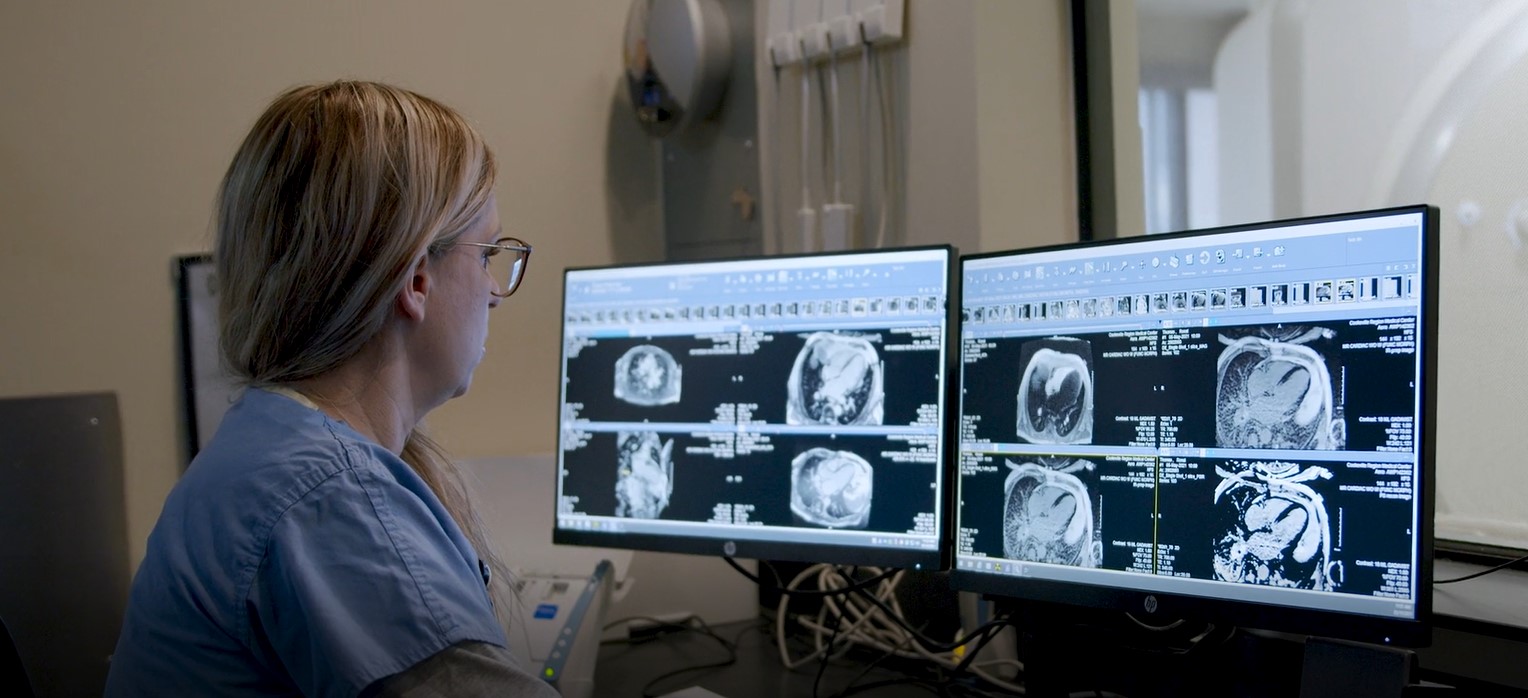
How will I get my results?
An interpreting physician (usually a radiologist) will read your exam and make a final diagnosis. His interpretation will be converted to a written report, which will be sent to the physician who ordered your exam. Your physician should present the findings of your exam to you. You may also sign up for a patient portal account at mycrmchealth.org to read your results online.
CONTACT US
931-783-2222
Cardiac CT
The heart is difficult to image because of its movement, but thanks to advances in CT technology, we are now able to take images of the heart with “frozen motion,” when the heart rate is reasonably low. This usually requires a pre-medication regimen prior to having this procedure.
Cardiac computed tomography (CT) refers to using CT to capture images of the heart and vessels connected to the heart. This may be performed with or without the use of intravenous contrast. Most applications require the use of contrast.
Cardiac CT evaluates the structure of the heart but it also can evaluate its function. Most heart diseases can be detected by a cardiac CT,, such as:
- Coronary artery disease, which is plaque in the coronary arteries. The CT can detect how it impacts the heart muscle.
- Atherosclerosis, which is the gradual clogging of the arteries by fatty materials and other substances in the blood stream. It develops over many years.
Cardiac CT is also used to guide procedures in cardiology, like ablation therapy for heart rhythm abnormalities or prior to heart valve replacement. Cardiac CT may be used to exclude a large blood clot in the cardiac chambers and prior to closure of the left atrial appendage.
HeartFlow Analysis
Coronary artery disease (CAD) is the leading cause of death for both men and women in the United States. CAD develops when the arteries leading to the heart narrow or become blocked, which may lead to a reduction in blood flow to the heart, causing chest pain, heart attacks and death.
Despite being the most common form of heart disease, many of the non-invasive tests available today have low accuracy rates in detecting the disease. Cookeville Regional Medical Center is advancing the diagnosis of CAD with HeartFlow Analysis. This non-invasive heart test provides a personalized 3D model of your coronary arteries that shows how each blockage impacts blood flow to your heart. This detailed information, which was previously only available through an invasive procedure, helps your doctors determine the next step in your treatment plan. This is used in conjunction with a standard coronary CT scan to provide a more detailed view of the significance of your coronary artery blockage.
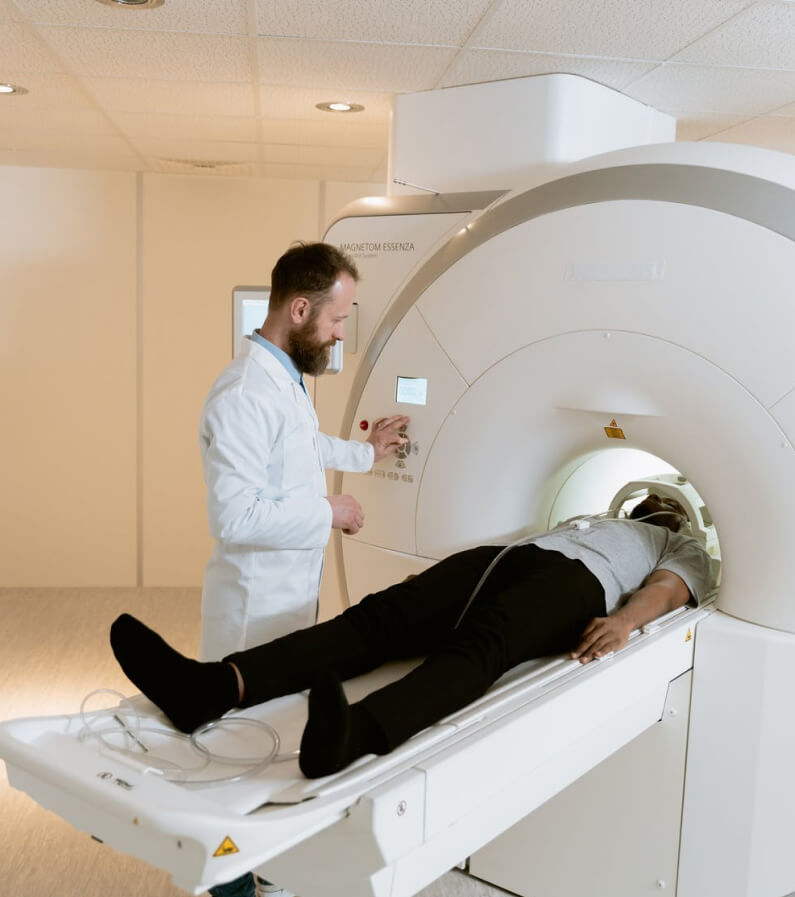
Cardiac MRI
Magnetic resonance imaging (MRI) may be used instead of a CT scan when organs or soft tissues are being studied.
MRI is a test that uses a large magnet, radio signals, and a computer to make images of organs and tissue in the body. In this case, the heart is imaged.
The MRI machine is large and tube-shaped. It creates a strong magnetic field around the body. Some MRI machines are more open.
Cardiac MRI may be done to assess signs or symptoms that may suggest:
- Cardiomyopathy. This happens when the heart muscle becomes thick and weakened.
- Myocarditis. Inflammation of the heart muscle.
- Congenital heart disease. These are defects in the heart that happen as the fetus forms. An example is a hole in the wall between the two lower chambers of the heart (ventricular septal defect).
- Heart failure. This condition means the heart muscle is weak and can’t pump enough blood to the body.
- Aneurysm. This is a widening and weakening of a part of the heart muscle or the aorta.
- Heart valve disease. When heart valves become damaged, it can block blood flow in the heart.
- Cardiac tumor. A tumor of the heart may happen on the outside surface or inside the heart.
There may be other reasons for your healthcare provider to recommend an MRI of the heart.
MRI cardiac stress testing
Cookeville Regional also offers MRI cardiac stress testing. This type of stress test is used to check the blood flow to the heart and also evaluate heart wall motion or abnormalities.
Echocardiography (cardiac ultrasound)
Echocardiography (ultrasound) is used to assess different areas of the heart and can detect heart disease or signs of serious cardiovascular conditions. Many factors contribute to an accurate diagnosis. The training and experience of the sonographer, the type of equipment used and the quality assessment metrics Cookeville Regional is required to measure, all contribute to a positive patient outcome.
Cookeville Regional Medical Center has been awarded accreditation by the Intersocietal Accreditation Commission (IAC) in Echocardiography for providing quality cardiac imaging.
Echo with contrast
An enhancement agent is administered through the vein when this type of Echo is done. The enhancement agent allows you to see the flow of blood and the heart muscle better.
Stress Echocardiogram
Also called stress echo, is a procedure that determines how well your heart and blood vessels are working. Your heart rhythm and blood pressure are monitored while you walk on a treadmill.
Dobutamine Stress Echo (DSE)
This type of stress test can be used if you are unable to exercise. Dobutamine is put through a vein and causes the heart to beat faster. It mimics the effects of exercise on the heart.
Bicycle stress echo
This type of stress echo involves a supine bike and allows the patient to pedal (exercise) while lying flat on the bed. The heart rhythm and blood pressure will also be monitored.
Transesophogeal Echo (TEE):
This type of Echo is done at the hospital. A cardiologist inserts a thin flexible tubing down the esophagus to obtain images of the heart. The TEE provides clearer and more detailed images of the left ventricle than a standard echo. Medication to make you relax will be given with this test.
Nuclear medicine
Myocardial Perfusion Imaging: Diagnosing coronary artery disease and accessing cardiac muscle as well as electrical activity.
MUGA scan: Evaluates how efficiently the heart functions as a pump and evaluates damage from cardiotoxic drugs or a heart attack.
Cardiac amylidosis scan: This detects the presence of harmful Amyoloid tissue in the heart that prevents unnecessary biopsies. Cookeville Regional is one of a few facilities that offers this procedure and is the only facilities between Nashville, Knoxville and Chattanooga.
PET myocardial viability scan: Evaluates the extent of cardiac muscle damaged from a heart attack, differentiates between hibernating/stunned heart muscle and true scarred muscle. This lets the cardiologist determine if revascularization would benefit the patient. Cookeville Regional is one of a few facilities that offers this procedure and is the only facility between Nashville, Knoxville and Chattanooga.